Recent research highlights the importance of developing and utilizing cost-effective, sensitive, and specific plasma biomarkers using commercially available assays for screening subjects in Alzheimer’s Disease (AD) clinical trials. This approach overcomes limitations associated with the clinical diagnosis of AD, especially in individuals with mild cognitive impairment or dementia who exhibit typical AD symptoms lacking Aβ pathology.

There is evidence that targeting p-tau217 in blood has yielded the best results as a diagnostic and prognostic tool for assessing longitudinal change. Studies showed that the p-tau217 assay outperformed MRI and showed comparable performance with CSF biomarkers in detecting Aβ PET positivity and tau PET positivity.

Utilizing the approach recommended by Alzheimer’s Association guidelines, commercial immunoassay has shown high positive and negative predictive accuracy at screening in clinical studies.

Learn about other advances in Alzheimer’s Disease research, including why the FDA’s recognition of using medical imaging as an objective biomarker is a big step forward for the clinical development of new AD treatments.

Rohit Sood PhD, VP Scientific & Medical Services, Calyx
Author:

Rohit Sood, MD, PhD, VP Scientific & Medical Services, Calyx

A Conversation with Oliver Bohnsack, MD, PhD, MBA

In February 2022 the Journal for ImmunoTherapy of Cancer published “Comparison of tumor assessments using RECIST 1.1 and irRECIST, and association with overall survival.” This publication marks the first time immune-related criteria show a correlation with overall survival as the most meaningful endpoint in the treatment of cancer patients.

Calyx’s Head of Oncology, Dr. Oliver Bohnsack is considered one of the leading industry experts on oncology-imaging trial design and response criteria, having co-authored the immune-related response criteria (irRC, 2009), authored irRECIST (2014), and co-authored Comparison of Assessments using RECIST and irRECIST by Eggleton P. et al. (2020).

Here we discuss with Dr. Bohnsack the implications of these new response assessments, based on his contribution as a co-author of the 2022 paper, and what they mean for oncology research and treatment decision-making moving forward.

 Watch Video


Watch this discussion with Dr. Oliver Bohnsack and Peter Eggleton on “Solid Tumor Trials: Maintain More Patients with irRECIST

“In my opinion irRECIST easily can and shall replace outdated RECIST 1.1 on all solid tumor trials going forward.”

– Dr. Oliver Bohnsack, VP, Medical Imaging & Head of Oncology, Calyx

Q: Can you give us some background on how cancer treatment response is currently measured in clinical trials?

Response Evaluation Criteria In Solid Tumors (RECIST) is a set of published rules that define when cancer patients improve (“respond”), stay the same (“stable”) or worsen (“progression”) during treatments. The original RECIST was published in February 2000 by an international collaboration and updated to RECIST 1.1 in 2009.

Today, most clinical trials with imaging-based response or progression as an endpoint evaluating cancer treatments in solid tumors are using RECIST 1.1.

Q: Are there limitations to using RECIST 1.1 for tumor assessment?

Yes, there are. Patients treated with immune checkpoint inhibitors (ICIs), or any other form of immune-based treatment may experience pseudoprogression, which shows – at least on imaging – a tumor burden worsening before the treatment shows efficacy in and is visible on scans. In this situation these patients will be classified as progressive disease (PD) by RECIST V.1.1 and thus could lead to inappropriate treatment discontinuation.

As a result, immune-response criteria – irRECIST (Immune-related Response Evaluation Criteria In Solid Tumors) ‒ were developed and used in many clinical trials since to better capture novel response patterns seen with immune-based treatments and T-cell therapies.

Q: How does irRECIST differ from RECIST 1.1?

The irRECIST approach allows responses not typically observed in traditional systemic treatment to be identified and better documented. The guideline describes a standard approach to solid tumor measurement and definitions for objective change in tumor size which can be used not only in immunotherapy clinical trials. Where any new lesion seen with RECIST 1.1 defines a treatment failure with documented PD, irRECIST allows for a possible continuation and further evaluation taking new lesions and the whole tumor burden into consideration.

Q. Can you tell us about the current findings outlined in the 2022 paper you contributed to?

Working with researchers from Merck Healthcare KGaA and renown clinical institutions we pooled data from 1765 patients with 12 types of advanced solid tumors treated with avelumab (an anti-programmed death ligand 1 (PD-L1) monoclonal antibody) monotherapy in the JAVELIN Solid Tumor and JAVELIN Merkel 200 trials, conducted a comparative analysis of tumor assessments by investigators according to RECIST 1.1 and irRECIST, and evaluated the correlation between progression-free survival (PFS), continued patient benefit and overall survival (OS).

The use of irRECIST identified a subset of patients with a best overall response (BOR) of progressive disease by RECIST 1.1 but an irBOR of immune-related disease control by irRECIST with a distinctive survival curve, thereby providing more clinically relevant information and better treatment decision-making options than RECIST 1.1 alone.

The publication demonstrates the benefit to a subgroup of patients in each of its various analyzed tumor indications, who otherwise would have foregone treatment and survival benefit when relying solely on RECIST 1.1 instead of irRECIST.

Q. What are the implications for sponsors of ICI trials who use RECIST 1.1 for evaluating response to treatment in studies of solid tumors?

Because immune-targeted treatment can initially cause the tumor burden to look as if it is progressing, when in fact it is not, clinical trial sponsors using RECIST 1.1 to assess treatment response may end up discontinuing patients who would otherwise remain in the study. Using RECIST 1.1 could limit sponsors from recognizing the full treatment benefit of new therapies in development, and more importantly, may prevent patients from receiving potentially beneficial treatments.

Q: What can we expect moving forward, as the industry considers these findings and their implications?

Sponsors and regulatory agencies will have to consider whether based on this data RECIST 1.1 is still appropriate to be used and advocated for as the current assessment standard for physicians, aiding them in treatment decision making and whether to continue or discontinue the current immune oncology treatment of their patients. irRECIST includes and covers all that’s embedded in RECIST 1.1 already but now takes the entire tumor burden including new tumor growth into consideration. In my opinion irRECIST easily can and shall replace outdated RECIST 1.1 on all solid tumor trials going forward.

The landscape of imaging in Myeloma patients has changed rapidly over the past few years. Routine clinical practice and clinical trial settings now utilize more advanced imaging modalities like PET-CT, whole-body MRI, and low-dose CT as compared to whole-body skeletal surveys (Xray) which were more commonly used in the past. These high-resolution imaging modalities lead to higher lesion detection rates at screening and thereby require an ongoing imaging evaluation in a predefined manner to support the efficacy analysis of a therapeutic intervention in clinical trials.

The figures below illustrate imaging presentation in myeloma patients and provide guidance on categorizing these lesions for response evaluation:

CT scans from a Myeloma Trial

Figure a) is a CT head of a patient showing a punched-out lytic lesion in the frontal bone (white arrow). Lytic lesions should be monitored qualitatively as per clinical discretion for progressive disease.

Soft tissue plasmacytoma noted in the pelvis (yellow arrows) in two different subjects: contrast-enhanced CT of pelvis (figure b) and a fused FDG PET image (figure c). Such soft tissue plasmacytoma need to be quantitatively evaluated at a predefined imaging schedule to evaluate the response of therapy along with the other lab parameters.

Regulators are looking for standardized methodologies to be applied upfront in trials to ensure the robustness of the data and its validity. We have noted a trend in the last year wherein health authorities across the globe are asking for a central review of imaging evaluation to mitigate variability in data from site imaging interpretation and across trials.

Choose Medical Imaging.


Learn how Calyx helped a leading pharmaceutical company receive accelerated approval for a Multiple Myeloma treatment.

Calyx is a leading provider of myeloma trial imaging, having supported nearly 40 trials and 6 approved indications to date. We routinely collaborate with myeloma trial sponsors and consult on how to standardize image acquisition and support independent, harmonized imaging analysis to meet regulators’ expectations.

Our expert methodology is based on the wealth of experience gained by supporting trials over the last 25 years and engaging in scientific consultation with experts such as Dr. Shaji Kumar and Dr. Joseph Mikhael.

Contact us to explore how our scientific and medical imaging experts can partner with your team to reduce risks and deliver reliable imaging data that meets regulators’ requests and sets your development program up for success.

2023 brought many healthcare advances and opportunities for life science professionals to stay current on the research, technologies, and processes that are driving change in how new medical treatments are developed and ultimately approved for worldwide use.

So here, in case you missed them, are the most sought-after articles, videos, case studies, and more produced by Calyx scientific, technical, and regulatory experts this year. Each provides direction and perspective on optimizing and accelerating the clinical development and approval of medical treatments.

We hope you find them as insightful and valuable to you now as they were the first time around.

De-risking Medical Imaging in Solid Tumor Trials

During anti-tumor treatment development, imaging modalities, criteria, and regulators’ expectations change frequently. Without the direction of imaging scientists who work day in and day out in clinical trial imaging, it would be difficult, if not impossible to keep track of and react to changes during these critical and often, lengthy trials.

Successful medical imaging in solid tumor trials requires professionals with therapeutic experience, expertise in the modalities required to demonstrate safety and efficacy, and first-hand insight into what global regulators will look for in your submissions.

This blog presents examples of scientific advances and regulatory changes that are currently impacting anti-tumor treatment research, demonstrating the need for an imaging partner who is immersed in the regulations, scientific learnings, and trends that could impact the success of your development program.

READ THE BLOG

Overcoming Oncology RTSM Challenges with Advanced IRT

Sponsors of oncology trials face unique randomization and trial supply management (RTSM) challenges, including:

  • Central vs. local sourcing of standard-of-care treatments
  • Unknown patient treatment duration
  • Impact of rescue medication on study drug expiration
  • High cost of treatments / need to reduce excessive drug wastage

Learn about these and other supply challenges and how a flexible IRT system can reduce errors, ensure patient safety, and meet changing RTSM during lengthy oncology trials in this article by Calyx’s Malcolm Morrissey, published in International Clinical Trials®.

READ THE ARTICLE

Exceeding Timelines for Accelerated Approval: Calyx Medical Imaging

In oncology clinical development, every day matters. Even more so when your compound has been fast-tracked for approval by global regulators.

Which is why so many clinical trial sponsors rely on Calyx Medical Imaging. Calyx’s collaborative approach, ability to meet each sponsor’s unique needs, and expertise in image acquisition and analysis have repeatedly been proven to help clinical development programs succeed.

Like in this example, where Calyx Medical Imaging delivered critical imaging data in advance of deadlines on an already expedited timeline to help a leading pharmaceutical company receive accelerated FDA approval for a Multiple Myeloma treatment.

WATCH THE VIDEO

How IRT Expertise Surfaces – and the Difference it Makes

An IRT system is a critical part of a clinical trial. The implications for failing to get it right can be impactful not only to study goals but to participants as well.

Here, Calyx’s Craig Mooney gives examples of the consequences that can arise if a study’s IRT system isn’t implemented with insight and precise focus on the protocol’s needs and why expertise matters throughout the clinical trial lifecycle.

READ THE BLOG

Calyx RIM Supports Successful FDA eCTD 4.0 Pilot

Calyx RIM has been successfully used by a global market-leading pharmaceutical company in FDA’s eCTD 4.0 implementation pilot program.

Calyx’s regulatory experts worked closely with this leading company and the FDA throughout the pilot and advanced Calyx RIM based on their feedback and lessons learned.

Learn why you can rely on Calyx RIM as you adopt eCTD 4.0 as part of your global regulatory processes.

READ THE ANNOUNCENENT

Neuroimaging in Alzheimer’s Disease Trials

As our understanding of Alzheimer’s Disease (AD) pathophysiology continues to evolve, we’re seeing advanced approaches for assessing treatment effects in clinical development, including AI/machine learning to measure subtle changes that are difficult for the human eye to detect.

Calyx Medical Imaging delivers neuroimaging expertise to help sponsors meet the unique and emerging needs of early to late-phase AD clinical trials. This paper outlines Calyx’s capabilities and experience, which includes confirmation of eligibility and brain safety assessments with rapid turn-around times as well as advanced quantitative analyses for PET and MRI data to help your AD trial succeed.

READ THE PAPER

Mitigate Unblinding Risks with Calyx IRT

Every clinical trial runs the risk of unintentional unblinding. With 30 years of experience designing reliable RTSM solutions and the processes behind it, Calyx IRT is the solution you can rely on to minimize these risks and ensure the integrity of your clinical trial.

The solution design and technical experts behind Calyx IRT have published a variety of papers, webinars, and blogs to demonstrate where and how unblinding can occur and more importantly, how to minimize those risks.

Access Here

Calyx CTMS Select: Scalable for Small/Mid-sized Biopharma and CROs

The features of Calyx CTMS deliver significant benefits for studies of all shapes and sizes. However, not all studies have the same needs.

Enter Calyx CTMS Select, a pre-configured and validated clinical trial management system for small to mid-tier biopharmaceutical companies and CROs who want reliability and global support but don’t require CTMS customization.

Calyx CTMS Select enables SMID organizations to scale as their operations require and includes the rich features required for clinical trial management oversight and monitoring at lower costs than comparable solutions.

READ THE ANNOUNCENENT

Cookie Cutter Solutions don’t Cut it in Clinical Trial Imaging

When medical imaging is used as a clinical trial biomarker, your centralized core lab provider needs therapeutic/modality and operational expertise to help you meet your development objectives, regardless of where you are in the clinical development spectrum.

Here we review how a small biotech captured the imaging data needed to secure funding and advance their early-stage research on a rare neurological disease compound. And, how a top 5 global pharma captured reliable primary efficacy endpoint data in a pivotal phase III study to achieve regulatory approval on a prostate cancer compound. All supported by Calyx Medical Imaging.

READ THE BLOG

During anti-tumor treatment development, imaging modalities, criteria, and regulators’ expectations change frequently. Without the direction of imaging scientists who work day in and day out in clinical trial imaging, it would be difficult, if not impossible to keep track of and react to changes during these critical and oftentimes, lengthy trials.

Successful medical imaging in solid tumor trials requires professionals with therapeutic experience, expertise in the modalities required to demonstrate safety and efficacy, and first-hand insight into what global regulators will look for in your submissions.

Here we present examples of scientific advances and regulatory changes that are currently impacting anti-tumor treatment research, demonstrating the need for an imaging partner who is immersed in the regulations, scientific learnings, and trends that could impact the success of your development program.

Example 1: Prostate Cancer

In prostate cancer research, PSMA-PET tracers are now commonly accepted for the detection of metastatic disease that was previously not visible on conventional imaging with CT/MRI and 99mTC Bone Scan. Patients and investigators are reluctant to perform conventional imaging with CT or MRI plus 99mTc bone scan when the more reliable and sensitive PSMA exams reveal more information about treatment efficacy or lack thereof.

Sponsors currently developing prostate cancer treatments are facing severe challenges in study design and meeting their protocol-defined endpoints based on conventional imaging alone. Even worse, as these pivotal trials take several years to complete, the modality and method of CT/MRI plus 99mTc Bone Scan may be outdated and no longer acceptable by the time the study closes.

In addition, there are several PSMA tracers available, they are similar yet not equal in their sensitivity and have slightly different uptake specifics. Plus, not all of these tracers are ready to be used in trials nor approved or available across different continents and countries.

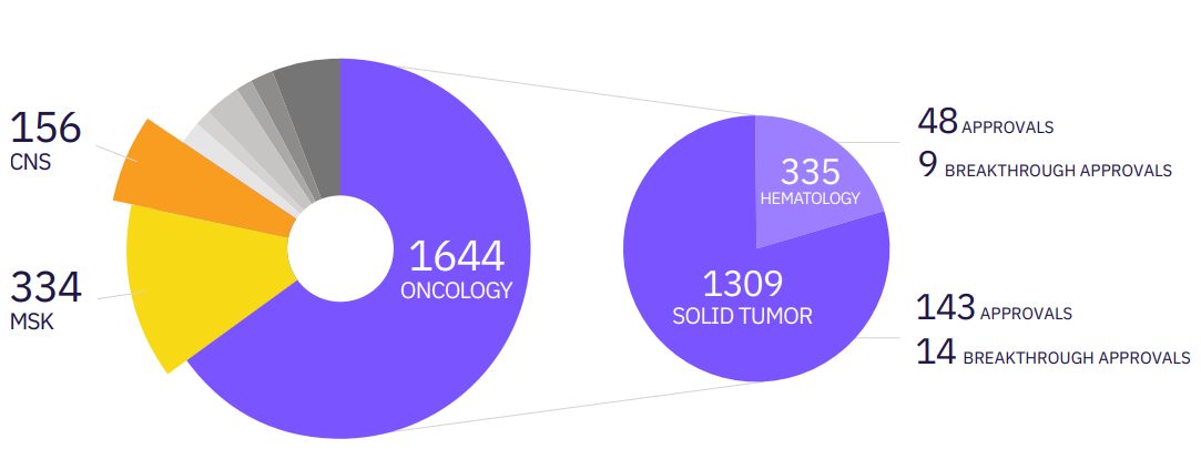
Calyx Medical Imaging has supported the approval of over 140 solid tumor treatment approvals, including this example of a phase III prostate cancer trial which led to the approval of a new treatment option for patients with mCRPC.

Example 2: Immuno-oncology research

Another example is related to the advances we’re seeing in immuno-oncology research. Recent findings are driving modifications to established imaging criteria, as evidenced in a Journal for ImmunoTherapy of Cancer paper (2022), marking the first time immune-related criteria show a correlation with overall survival as the most meaningful endpoint in the treatment of cancer patients.

A recent paper demonstrates irRECIST may provide more clinically relevant information and better treatment decision-making options than RECIST 1.1 alone in solid tumor image assessment.

Because immune-targeted treatment can initially cause a tumor to look as if it is progressing when it is not, clinical trial sponsors using RECIST 1.1 to assess treatment response may end up discontinuing patients who would otherwise remain in the study. Based on data from 1765 patients with 12 types of advanced solid tumors treated with avelumab monotherapy, the study found that irRECIST may provide more clinically relevant information and better treatment decision-making options than RECIST 1.1 alone in solid tumor image assessment.

As a result, Dr. Oliver Bohnsack (see sidebar) suggests that irRECIST can and should replace outdated RECIST 1.1 on all solid tumor trials going forward.


Calyx’s Dr. Oliver Bohnsack leverages his knowledge from having co-authored the immune-related response criteria (irRC, 2009), served as first author of irRECIST (2014), and co-authored Comparison of Assessments using RECIST and irRECIST by Manitz J. et al. (2020) as he designs and implements optimal imaging strategies for Calyx’s customers developing new oncology treatments

Solid Tumor Trials: Maintain More Patients with irRECIST

Calyx Medical Imaging extends groundbreaking capability from Ceretype to improve psychiatric treatment outcomes

Nottingham, England and Morrisville, NC – April 4, 2023 Calyx, the eClinical and Regulatory solutions and services provider relied on for solving complex data challenges in clinical research, today announced a strategic partnership with Ceretype Neuromedicine, Inc., whose novel functional MRI (fMRI) platform incorporates optimal image acquisition and advanced analytics to de-risk and accelerate the development of psychiatric and neurological therapies.

The current lack of objective biomarkers presents a real challenge for researchers developing treatments for depression/anxiety, schizophrenia, and other neurological/psychiatric conditions such as Parkinson’s Disease. An in-market, audit-cleared “biomarker engine,” the Ceretype platform utilizes cutting-edge fMRI and artificial intelligence (AI) to connect neural circuitry to patient behaviors and treatment outcomes, delivering the objective biologic endpoints researchers need for more precise measurement of treatment effects during central nervous system (CNS) drug development.

“A key component of Calyx’s strategy is partnering with best-in-class technology providers to offer the innovative imaging biomarkers required to find new treatments for unmet medical needs,” said Stephen M. Bravo, MD, Chief Medical Officer at Calyx. “We’re excited to partner with Ceretype and for our customers to leverage their advanced fMRI technology to bring new and more effective psychiatric treatments to market.”

“We’re delighted to combine Ceretype’s neuroscience, biophysics, and computing expertise with Calyx’s 25 years of experience delivering reliable clinical trial imaging,” said Emily Stern, CEO at Ceretype. “Through this partnership, we will achieve our mission of improving psychiatric and neurologic patient outcomes through precision drug development and precision treatment.”

For more information visit Calyx.ai/Imaging and ceretype.com.

About Calyx

Through innovative eClinical and Regulatory solutions and services, Calyx turns the uncertain into the reliable, helping bring new medical treatments to market reliably. With deep expertise in clinical development and 30 years supporting trial sponsors and clinical research organizations, Calyx harnesses its intelligence and experience to solve complex problems, deliver fast insights, and get new drugs to market every day.

CTMS | EDC | IRT | Medical Imaging | RIM

Take your trials further with intelligent insights at Calyx.ai or at LinkedIn, Twitter, or Facebook.

About Ceretype, Inc.

Ceretype provides actionable insights on brain function – both for individuals and for populations. Our platform de-risks and accelerates clinical trials targeting therapeutics for the central nervous system.  Whole-brain, circuit-level, actionable insights, (e.g., identification of mechanism of action, target engagement, positive response profiles) are delivered across all trial phases, for any intervention and for any neurological or psychiatric disease.  In addition to clinical trials, Ceretype will bring our technology and treatment-specific biomarkers to patients for biologically informed, personalized patient care.

Christine Tobin | Christine.Tobin@Calyx.ai | +1 412-628-8598

Stay Updated

Never miss a beat. Sign up to receive emails covering industry news and useful content to help you advance clinical development.

  • This field is for validation purposes and should be left unchanged.