When medical imaging is used as a clinical trial biomarker, it’s critical that you select a centralized core lab provider whose experience, capabilities, and approach to medical imaging will help you meet your development objectives, whatever they are.

This applies whether you’re a small biotech needing reliable imaging data to demonstrate your compound’s potential as you seek funding to advance your research and/or to license it to a development partner. Or if you’re one of the world’s largest pharmaceutical companies needing data from a pivotal phase III study to demonstrate safety and efficacy as you seek regulatory approval. And every scenario in between.

Regardless of your objectives, when imaging data will make the difference between success and failure, you can trust Calyx Medical Imaging to help you succeed. Here we present different scenarios in which the scientific expertise, operational experience, and dedication of Calyx’s Medical Imaging team helped researchers on completely opposite ends of the clinical development spectrum achieve their development goals.

Case Study 1:

Small biotech – Early-stage research on rare neurological disease compound

A small biotech company was at the very early stages of investigating a compound for a rare neurological disease for which no approved treatments were available. The company needed an imaging partner with neurological expertise to help them characterize and determine the best modality for measuring disease progression and, eventually, the compound’s efficacy.

After talking to other imaging providers and finding their ‘cookie cutter’ approach not suitable to their research objectives, the company chose to partner with Calyx. The company valued Calyx’s expertise and ability to offer the customized services they needed to move forward in their research.

The relationship began with collaboration between Calyx Medical Imaging experts and the company’s scientific and medical team in compiling and reviewing all available data on the disease presentation, focusing on how medical imaging (MRI, CT) and pathology had been used to diagnose and track disease progression to date.

Calyx scientists then developed a briefing book which presented two imaging-based models of the disease progression and the role medical imaging would play in drug development. The briefing book was ‘FDA ready’ and as such, the company received regulatory approval to move forward.

Continuing the partnership on research of the rare disease, the company contracted Calyx for a natural history study to collect data on the disease presentation and progression for future development of novel therapies. Magnetic Resonance Imaging (MRI) was utilized to develop and evaluate imaging-based biomarkers for assessing the burden and tracking the natural history of this rare disease.

The findings from the natural history study guided and informed the client on the imaging-supported endpoints to be used in their Phase 2 clinical trial on a therapy/compound for the rare disease.

The company continued to rely on Calyx for the design and execution of the phase 2 and natural history studies, which involved Calyx’s standard imaging services and operations, including the development of an independent review charter, training of site personnel on image acquisition, and selection / training of neuroradiologists. In addition to performing thorough quality checks of images captured in the study, Calyx’s Medical Imaging team quickly responded to questions that arose during imaging reads and took an additional, hands-on approach with reviewers and the company’s scientists to ensure the quality of the read results.

During an investors’ event the company shared the promising data – the majority of which were the first to characterize the disease state – as well as updates on the natural history study and their plans to develop the compound further. The data and the rapid progress of the ongoing study were well received by the investors, which enabled the company to continue its research. In the word’s of the company’s VP, Clinical Development Sciences, “We could not have achieved this important milestone without Calyx’s support.”

Case Study 2:

Top five pharma – Phase III trial of prostate cancer treatment

One of the world’s top five pharmaceutical companies was in the late stages of development for a new treatment for metastatic, castration-resistant prostate cancer (mCRPC). Because this advanced form of prostate cancer doesn’t respond well to currently available treatments, mCRPC presents itself particularly challenging for patients and medical professionals alike.

The company selected Calyx Medical Imaging to support its pivotal Phase III trial, which enrolled over 900 patients from over 150 worldwide investigative sites. The imaging data collected throughout the four-year study would support the sponsor’s primary efficacy endpoint and be included in their submission for EU regulatory approval.

For this study, Calyx designed and delivered robust Medical Imaging services that included:

A comprehensive imaging charter, following the Prostate Cancer Working Group 3 (PCWG3) criteria, developed by Calyx’s professional medical writing team.

  • Proven, scalable services to drive the collection of CTs, MRIs, and bone scans, which involved:
    • Training of investigative sites on the correct image capture procedures
    • Collection and quality review of over 22,000 CTs, MRIs, and Bone Scans
  • Advanced internal processes to rule out false positives in bone scans
  • Blinded Independent Central Review (BICR) of imaging reads by 20 expert reviewers
  • A dedicated scientific/medical team with expertise across multiple oncology indications and imaging modalities

Calyx’s scientific and medical team (SciMed) remained intact over the four-year study, consulting with the sponsor and supporting the investigative sites and reviewers throughout to ensure the data derived from the important imaging analysis was of the highest quality.

Calyx met all the deliverable dates throughout the study and supplied reliable imaging data to support the study’s primary efficacy endpoint, demonstrating that the treatment significantly reduced the risk of disease progression or death compared to traditional treatment approaches.

As a result of this study the robust and reliable data supplied by Calyx Medical Imaging, the new and better treatment was approved in the EU and is now giving patients a safe and more effective treatment option for mCRPC.

Related

Photo taken in Helsinki, Finland

Medical Imaging in Rare Disease Trials: Idiopathic Pulmonary Fibrosis (IPF)

Image 177

Managing a Breakthrough: Navigating the Operational Challenges of Breakthrough Trial Imaging

RDIblog-MI-header2000x1000_v1

Partnering for Rare Disease Clinical Trial Success

2022 brought many healthcare advances and opportunities for life science professionals to stay up to date on the research, technologies, and processes that are driving change in how new medical treatments are developed and ultimately approved for worldwide use.

So here, in case you missed them, are the most sought-after articles, white papers, webinars, and more produced by Calyx scientific, technical, and regulatory experts this year. Each provides direction and perspective on optimizing and accelerating the clinical development and approval of medical treatments. We hope you find them as insightful and valuable to you now as they were the first time around.

This blog series addresses the trigger points for unintentional unblinding, how partial unblinding can become full unblinding, and some controversial issues related to unintentional and partial unblinding in clinical trials.

In 2021, Calyx advanced its strategy of partnering with best-in-class technology providers to offer innovative imaging biomarkers required to find new treatments for unmet medical needs. Our groundbreaking partnership with Qynapse enables our clients to more confidently assess the full potential of treatments in development for Multiple Sclerosis, Parkinson’s, Alzheimer’s, and Huntington’s disease, as well as other neurodegenerative disorders. And our partnership with Neosoma delivers novel, improved AI-based neuro-oncology imaging assessment to clinical trial sponsors developing new treatments for gliobastoma and other life-threatening neuro-oncological diseases.

The Journal for ImmunoTherapy of Cancer recently published ‘Comparison of tumor assessments using RECIST 1.1 and irRECIST, and association with overall survival,’ marking the first time immune-related criteria show correlation with Overall Survival as its most meaningful endpoint in the treatment of cancer patients.

The publication demonstrates the benefit to a subgroup of patients who otherwise would have foregone treatment and survival benefit when relying solely on RECIST 1.1 instead of irRECIST, as irRECIST takes the entire tumor burden including new tumor growth into consideration.

In this webinar, co-authors Peter Eggleton of Merck and Oliver Bohnsack of Calyx – leading experts on RECIST and irRECIST – discuss the implications of these findings, what it means for oncology clinical development and treatment decision-making, and why irRECIST easily can and shall replace outdated RECIST 1.1 on all solid tumor trials going forward.

In rare disease trials, it is critical that trial supplies are available when patients are identified, and that drug overage is minimized – a real challenge due to larger numbers of investigative sites typically required to meet trial enrollment levels. Which is why we’re honored that so many sponsors of orphan drug-designated trials have entrusted their RTSM needs to Calyx IRT and continue to rely on our expertise as they bring safe and effective treatments to patients in critical need.  Learn more about Calyx IRT.

This blog series reviews the factors regulatory affairs professionals should consider as they prepare for eCTD 4.0 implementation, beginning with people and business processes, the impacts of new concepts and terminologies, and the challenges to be expected.

What are the consequences of deferring IRT functionality to meet clinical trial start dates? In this Applied Clinical Trials article, Calyx’s Craig Mooney reviews the inefficiencies and potential quality / regulatory risks likely to occur.

At Calyx, we have a long history as part of a CRO, so we know how a CRO functions, the challenges you’re up against, and the importance of keeping your customers satisfied. This inside knowledge affects every aspect of our CRO relationships and makes us more efficient and easier to work with.

Our Activate Solutions for CRO Partners Program enables CROs of all shapes and sizes to partner with Calyx and extend our advanced technology and proven services to drive success for their customers. In 2022 we added more CROs to the program – get to know some of them and learn how they’re partnering with Calyx to deliver more value.

80 sites enrolling 150 patients: How do you minimize overage without burdening your supply team? Learn how one of Calyx IRT’s advanced trial management options solved the complex in this case study.

In this episode of the Calyx Café, Patient Advocate Emily Epstein discusses the importance of supporting clinical trial patients’ mental health and the impact that clinical trial technologies – including DCTs – may have on patient welfare.

Your clinical trials deserve every chance to succeed. And your monitors deserve the most effective tools to improve trial efficiencies. This brochure describes why Calyx CTMS is the solution.

Partnership with Neosoma enables faster, more precise tumor assessment in glioblastoma research.

Nottingham, England and Morrisville, NC – November 17, 2022 – Calyx, the eClinical and Regulatory solutions and services provider relied on for solving complex data challenges in clinical research, today announced a strategic partnership with Neosoma, Inc., an innovative medical technology company focused on advancing brain cancer treatment. Through this partnership, Calyx Medical Imaging extends Neosoma HGG (High-Grade Glioma), a novel AI-based neuro-oncology software device to its worldwide customers who are developing treatments for glioblastoma, the most common and challenging, malignant primary brain cancer.

“Neosoma’s expertise in AI imaging development nicely complements Calyx’s two decades of experience in clinical trial imaging and the insights we’ve gleaned from having successfully supported nearly 100 neuro-oncology clinical trials,” said Stephen Bravo, MD, Chief Medical Officer, Calyx. “We’re pleased to extend Neosoma’s innovation to clinical development and give our customers greater confidence that the important imaging data they depend on to evaluate their glioblastoma compounds’ effectiveness is accurate and reliable.”

Clinical trials of glioblastoma treatments are complex, largely because gliomas have highly irregular and infiltrating shapes that are difficult even for experienced neuro-radiologists to delineate. Volumetric tumor assessments – which are becoming commonly used as exploratory endpoints – currently need to be conducted manually by neuroradiologists, which presents a time-consuming and difficult challenge for glioma image analysis in clinical trials. Leveraging advanced artificial intelligence, Neosoma HGG has been shown to achieve 95.5% accuracy in tumor volume measurement as established by consensus reads, which exceeds that of individual neuro-radiology experts and can save time while generating consistent, automated volumetric assessments of tumor change.

“Calyx’s Medical Imaging team is widely recognized as the leader in clinical trial imaging, especially in oncology, where experience and deep therapeutic area expertise are critical,” said Aly Abayazeed, MD, Chief Medical Officer, and Co-founder, Neosoma. ‘We are delighted to provide faster and more precise assessment of high-grade gliomas to their customers to advance glioblastoma trials and ultimately bring improved treatment options to patients with this life-threatening disease.”

This partnership will extend to other AI-based solutions being developed by Neosoma and reflects Calyx’s strategy of partnering with best-in-class technology providers to offer innovative imaging biomarkers required to find new treatments for unmet medical needs.

For more information on Calyx Medical Imaging, visit Calyx.ai/Imaging

About Calyx

Through innovative eClinical and Regulatory solutions and services, Calyx turns the uncertain into the reliable, helping bring new medical treatments to market reliably. With deep expertise in clinical development and 30 years supporting trial sponsors and clinical research organizations, Calyx harnesses its intelligence and experience to solve complex problems, deliver fast insights, and get new drugs to market every day.

Medical Imaging | IRT | CTMS | EDC | RIM

Take your trials further with intelligent insights at Calyx.ai or at LinkedIn, Twitter, or Facebook.

Contact:

Christine Tobin | Christine.Tobin@Calyx.ai | +1 412-628-8598

A lot has changed in how new treatments are developed for Alzheimer’s Disease (AD). With the 2021 approval of Biogen’s Aduhelm™, the FDA has clearly demonstrated a willingness to use imaging biomarkers as primary endpoints in AD, something that has never occurred before in the CNS space.

Here we talk with Calyx’s Dr. Stephen M. Bravo and Dr. Rohit Sood about how the use of medical imaging is advancing AD research and what we can expect moving forward.

Rohit Sood PhD, VP Scientific & Medical Services, Calyx

“As the technology and our understanding of AD pathophysiology evolves, we expect to see advanced approaches for assessing treatment effect in clinical development, ultimately enabling more effective Alzheimer’s Disease treatments.”

– Rohit Sood, MD, PhD, VP Scientific & Medical Services, Calyx

How is the research behind AD treatment development evolving?

We as an industry are still trying to understand the pathophysiology of Alzheimer’s. Although we’re continuously updating our knowledge based on new information coming out of the labs, it hasn’t yet been appropriately delineated.

Over the last decade our understanding was that beta amyloid was the primary protein responsible for the underlying pathophysiology of AD. Although the contribution of abnormal protein deposition to AD is widely recognized and currently accepted by the scientific community, the exact pathogenesis of AD is complex, and more effort needs to be put into understanding the pathophysiology of AD for the development of therapeutic agents. Moreover, as pharmaceutical clinical trials tailored toward the removal of beta amyloid have failed to demonstrate significant response, there is increasing interest in the role of Tau protein aggregates and beta amyloid plaque, with an emphasis on anti-Tau antibodies.

In addition, there is a school of thought that neuroinflammation plays a role and today, industry is in the process of developing radioisotope tags so that we can identify neuroinflammation and whether there’s a change in the extent of neuroinflammation with drug intervention.

All of these are important factors for trial sponsors to consider when deciding what to target in their AD development programs. But the decision on how to best assess treatment response is not always clear cut. There is an increasingly complex array of potential digital radiology biomarkers that look at brain perfusion and diffusion changes, and volumetric analysis of the hippocampus and other limbic regions including amygdala and entorhinal cortex.

As the technology and our understanding of AD pathophysiology continues to evolve, we expect to see advanced approaches for assessing AD treatment effect in clinical development, ultimately enabling the availability of more effective Alzheimer’s Disease treatments.

“Sponsors and CROs should partner with a core imaging lab whose scientists are experienced in AD study design and understand the optimal imaging modalities needed to demonstrate drug efficacy efficiently, effectively, and economically.”

– Stephen M. Bravo, MD, Chief Medical Officer, Calyx

Is medical imaging currently required in AD studies?

Neuromaging modalities such as PET and MRI have been used to detect pathological changes associated with AD and have played an important role in the development of noninvasive biomarkers of disease progression and assessment of response to therapy in clinical trials. Detection of early neurodegenerative changes associated with AD has been possible due to the development of neuroimaging measures of brain atrophy and brain structures such as hippocampus and cortex. Novel image analysis algorithms for characterizing and classifying AD and Mild Cognitive Impairment (MCI) have been developed and applied to MR images. MRI is also currently used for safety assessment of AD drug therapies, especially important in the development of monoclonal antibody therapies, which can cause life threatening changes in the brain. But now medical imaging is also going to be a requisite for primary efficacy endpoint data collection in order for FDA to consider new drug approvals.

What’s changing in how we assess drug efficacy in AD clinical trials?

Traditionally, efficacy in AD trials is evaluated via rater analysis by clinical doctors who are considered experts in the field of dementia – as well as geriatric, psychiatric, and neurology experts – using the mini mental status examination or other clinical tools to determine baseline disease level and progression or regression. Because these tools are very subjective and produce tremendous amounts of interobserver variability, rater analysis is becoming outdated and, while it will likely never disappear, will likely become less relevant in future trials.

The utilization of radiology biomarker data removes this subjective analytic bias and is a far more rigorous scientific methodology to determine drug effects. Currently, there are AI/machine learning techniques that are being developed to measure subtle changes that are difficult for the human eye to detect, delivering more perspective and a more scientifically rigorous data set that stands up to statistical scrutiny in determining treatment response.

How can Calyx help sponsors leverage medical imaging to demonstrate AD treatment efficacy?

Calyx’s Medical Imaging scientists have significant experience in Alzheimer’s Disease drug development and broad knowledge of the emerging modalities to improve drug response evaluation. They routinely help AD researchers:

1. Determine the best radiological biomarker to evaluate
2. Identify how the radiology modalities used can help to drive identification of an enriched study subject population yielding greater rates of enrollment/randomization
3. Identify digital radiological biomarkers which can be quantified and tracked longitudinally to provide more scientifically rigorous data supporting drug efficacy to the FDA
4. Produce reliable data to support regulatory submissions through the Blinded Independent Central Review (BICR) of imaging reads

What do you see in the future re: the impact medical imaging will have on AD treatment development?

As our understanding of AD advances, so does the technology that we’ll use to develop more effective treatments for it.  We see medical imaging delivering additional value in two ways:

  • Validating the companion diagnostics required to identify patients who will be best served by treatment of the drug being trialed. This drives the economic rational/model which allows for CMS (Medicare/Medicaid) and commercial US insurance reimbursement decisions.
  • Driving further quantitative analysis of MR and PET/CT images to derive even more sophisticated biomarker analysis to help determine the velocity of drug effects and to predict drug effect on an individual basis (supporting the notion of individualized medicine being applied to the clinical research/life sciences space).

Conclusion

FDA’s recognition of using imaging as an objective, noninvasive biomarker is a big step forward for Alzheimer’s Disease research. Clinical trial sponsors should partner with a core imaging lab whose medical imaging scientists are experienced in AD study design and understand the optimal imaging modalities needed to demonstrate drug efficacy efficiently, effectively, and economically.

Partnership delivers advanced approaches for bringing neurodegenerative disease treatments to market

Nottingham, England, Morrisville, NC, and Boston, MA–July 25, 2022 Calyx, the eClinical and Regulatory solutions and services provider relied on for solving complex data challenges in clinical research, today announced it has entered into a partnership with Qynapse, an Artificial Intelligence (AI) neuroimaging medical technology company. The partnership enables Calyx to adopt and deliver Qynapse’s precise and objective AI-powered neuroimaging analysis solutions, QyScore® and QyPredict® to its global pharmaceutical and biotech customers as they develop new medical treatments for central nervous system (CNS) disorders.

“We’re delighted to add Qynapse’s advanced analysis capabilities to our full suite of proven medical imaging services,” said Stephen M. Bravo, MD, Chief Medical Officer at Calyx. “This groundbreaking services and technology partnership will enable our clients to more confidently assess the full potential of treatments in development for Multiple Sclerosis, Parkinson’s, Alzheimer’s, and Huntington’s disease, as well as other neurodegenerative disorders.”

Qynapse has developed one of the largest datasets of proprietary algorithms designed to rapidly analyze and interpret brain scans, delivering more objectivity and precision for the measurement of drug efficacy and safety during CNS clinical trials. Calyx’s Medical Imaging services have been proven in over 2,600 global clinical trials, delivering reliable data that has supported the approval of over 270 new medical treatments.

“We are excited to partner with Calyx, a Medical Imaging and eClinical solutions powerhouse, in bringing life-changing treatments to patients globally,” said Olivier Courrèges, Chief Executive Officer at Qynapse. “Working together we will further expedite and improve the go-to-market process for CNS drug therapies.”

Click here for more information on Calyx’s expertise in CNS clinical trial imaging.

About Calyx

Through innovative eClinical and Regulatory solutions and services, Calyx turns the uncertain into the reliable, helping bring new medical treatments to market reliably. With deep expertise in clinical development and 30 years supporting trial sponsors and clinical research organizations, Calyx harnesses its intelligence and experience to solve complex problems, deliver fast insights, and get new drugs to market every day.

Medical Imaging | IRT | CTMS | EDC | RIM

Take your trials further with intelligent insights at Calyx.ai or at LinkedIn, Twitter, or Facebook.

About Qynapse

Qynapse is a medical technology company with an AI-powered and proprietary neuroimaging software platform that creates the potential for earlier clinical precision on the frontlines of CNS diseases. Qynapse’s flagship solution, QyScore®, FDA-Cleared and CE marked, combines MRI scans and AI to produce rapid, actionable insights for objective brain scan analysis, enhancing diagnosis precision and drug efficacy assessment. Qynapse’s prognostic AI technology, QyPredict®, available for research use only, has the potential to predict disease trajectory and improve targeted patient selection in clinical trials. Qynapse is headquartered in France, in the United States and in Canada.

Learn more about Qynapse’s AI solutions at https://qynapse.com/, or at LinkedIn, Twitter.

Contact:

Christine Tobin | Christine.Tobin@Calyx.ai | +1 412-628-8598

Reader fatigue in radiology can lead to high medical errors. Out of more than 11,000 preventable hospital deaths in English NHS acute hospitals each year, over eight thousand deaths are attributed to diagnostic error.

Interpretation of medical images is a challenging task as it is repetitive in nature, and errors (false negatives) are relatively common. Radiologists are also subject to visual fatigue due to continuous and longtime exposure to computer screens for interpreting medical images. It has been found that radiological errors are more likely to occur at the end of shift as compared to the beginning.

Blinded Independent Central Review (BICR) is one method recommended by the FDA for registration oncology trials (1). The objective evaluation of clinical indicators involving radiological images is an important end-point factor in oncology trials. During BICR, readers are routinely monitored using different models to ensure quality reads.

“Reviewer Disagreement Index may be used as an early indicator for potential reader quality and fatigue if a particular reader is concurrently reading on multiple trials.”

– Manish Sharma, MD, VP Medical Imaging, Calyx

Reader Monitoring Approaches

Monitoring of reviewers is not just mandatory from a regulatory viewpoint but also critical for proactive intervention throughout the trial. Monitoring safeguards high quality radiological assessments as well as adherence to specific clinical trial protocols. There is always inherent variability in the BICR process due to the difference in backgrounds, training, and humanity of the reviewers. Despite the existing inherent variability, there is a lack of meaningful methods for tracking and proactively improving reviewer performance.

“Double read with adjudication” is one of the frequently used models in various oncology trials review. This method permits better radiological assessments than a single read model. Double read with adjudication makes the process of BICR robust enough to not let variability and even minor errors impact the study outcome, if monitored properly.

Adjudication Rate (AR) has been used as a metric to track reviewer performance but does not accurately identify reviewer performance issues. It is dependent on many external variables like adjudication trigger, end point, indication, tumor burden etc. Adjudication agreement rate (AAR) is a relative performance indicator for a given reviewer where a higher adjudicator agreement rate suggests better reader performance.

Evaluating Reader Fatigue with Reviewer Disagreement Index

In an earlier blog, we presented how Reviewer disagreement index (RDI) considers the subjects for which adjudicator disagreed with the reviewer and considers adjudicator disagreement relative to the total number of cases read. RDI considers both the overall AR for a study and the individual AAR for each reader. The RDI indicates the percentage of disagreed cases for a given reader across the total number of cases read, as defined in the equation below, where a low RDI value indicates better reader performance and high RDI value indicates poor reader performance.

RDI =
# of cases where adjudicator disagreed with given reader
Total # of all cases read

Not surprisingly, there might be a correlation between the number of studies a reader reads on and how the cases are distributed across all studies. If the cases are distributed across too many studies, it can be a major issue contributing to reader fatigue e.g., if the reader in a reading session of 2 hours reads a few cases each on 10 different trials. The best-case scenario is to have a fine balance that would allow a reader a majority of cases on the same study in a reading session.

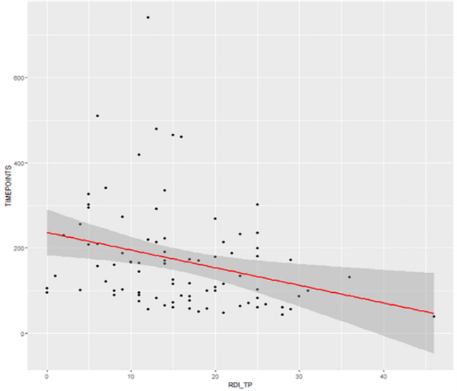
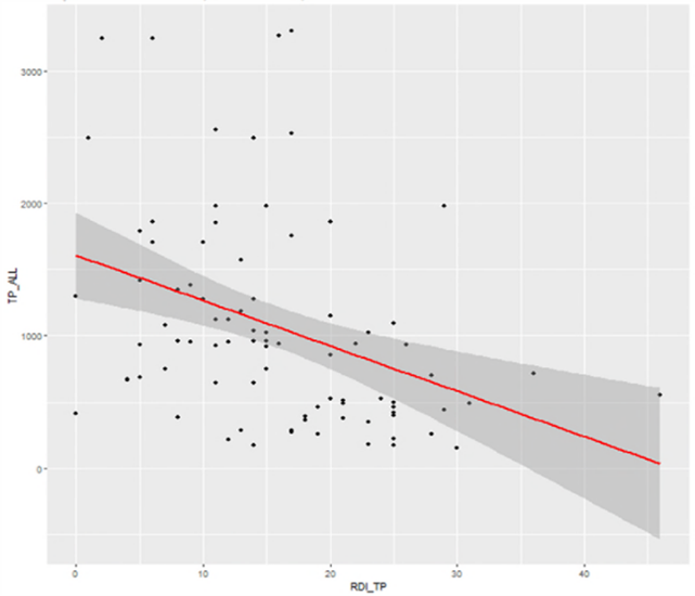
The plots below show a weak correlation of decreasing timepoints (Y-axis) to increasing RDI (X-axis). RDI can be used to give an early insight to trends by monitoring the ratio of RDI on each study per week, or per reading session and its interplay with overall and other studies’ RDI.

A recent study suggests that RDI can also be used as a good surrogate for reader fatigue (3). RDI may be used on an effective daily / weekly / monthly basis against read volume for monitoring reader fatigue which affects variability and thus read quality. Increasing RDI trend may be used as an early indicator for potential reader quality and fatigue if a particular reader is concurrently reading on multiple trials.

Reference

1. Guidance for Industry Developing Medical Imaging Drug and Biologic Products. Part 3: Design, Analysis, and Interpretation of Clinical Studies. US Department of Health and Human Services. Food and Drug Administration. Center for Drug Evaluation and Research. Center for Biologics Evaluation and Research; 2004.

2. Clinical Trial Imaging Endpoints Process Standards Guidance for Industry Draft. US Department of Health and Human Services. Food and Drug Administration. Center for Drug Evaluation and Research. Center for Biologics Evaluation and Research. March 2015 Revision1.

3. Manish Sharma, Madhuri Madasu, Sree Sudha Kota, Surabhi Bajpai, Yibin Shao, Srinivas Pasupuleti, Michael O’Connor, “Using reader disagreement index as a tool for monitoring impact on read quality due to reader fatigue in central reviewers,” Proc. SPIE 12035, Medical Imaging 2022: Image Perception, Observer Performance, and Technology Assessment, 120350J (4 April 2022); doi: 10.1117/12.2613082

Stay Updated

Never miss a beat. Sign up to receive emails covering industry news and useful content to help you advance clinical development.

  • This field is for validation purposes and should be left unchanged.